
A dental implant is a “root” device, made of titanium which supports restorations that resemble a tooth or group of teeth and the main function is to replace missing teeth.
They appear similar to an actual tooth root and are placed within the bone. The bone of the jaw accepts and fuses with the implant.
Single implant placement in areas of well-healed bone can often require no downtime, with patients returning to routine activity immediately. Some mild pain can be expected as this is associated with the healing process.

Why use 3D X-ray planning and not conventional xrays when planning dental implants?
Cone beam computed tomography (CBCT) is an essential tool for treatment planning and post-procedure monitoring. It provides highly accurate 3-D images of the patient’s anatomy from a single, low-radiation scan. CBCT technology delivers a comprehensive understanding of the patient’s jaw and the anatomical structures necessary to properly provide treatment.
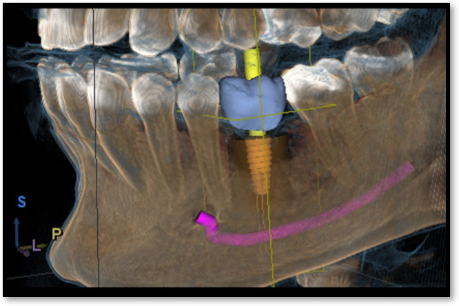
To ensure a successful implant placement, one must take into account the patient’s prosthetic needs, functional requirements, and anatomical constraints. During the assessments, 3-D imaging contributes to a greater success rate due to its ability to visualize previously undetectable anatomic variability and pathology.
CBCT-generated images provide detailed information that normal 2-D radiographs cannot offer. This greatly aids in precision, which improves the entire implant process.
Without the aid of 3-D imaging, dentists must speculate on bone density or exactly how deep implants need to be. CBCT images have less distortion than the 2-D images, and hence are more accurate in showing bone height and width. This allows for accurate measurements of jawbone thickness, while defining the shape of the bone contours.
One CBCT scan can include the entire maxillofacial region, including the upper jaw, lower jaw, base of skull, and joints. As a result, CBCT allows dentists to accurately locate the position of the nerve canal in the lower jaw, thus minimizing the risk of nerve damage. In the upper jaw, use of 3-D imaging during the implant planning process or surgical phase can aid inserting implants around the maxillary sinus, or assist in surgery where the maxillary sinus is augmented with bone.
What is Osstell Technology and why is it so important?
Patients want a nice smile and they want it sooner rather than later. More and more patients are asking for early and immediate loading of their implants, and patients who in the past might not have been candidates for implants are also asking to be treated. Patients request for shorter treatment times. There is an increasing need to evaluate implant stability and osseointegration (integration of the implant with bone) that cannot be achieved using traditional methods such as torque and percussion tests. These expectations require a more advanced diagnostics tool.
Monitoring osseointegration is critical to predictable restorative protocols. Using Resonance Frequency Analysis (RFA) technology, Osstell ISQ is the only objective, reliable and non-invasive way to monitor osseointegration of the implant. Osstell uses a universal ISQ scale, so a dentist can measure initial mechanical stability when the implant is placed, and have a baseline value so a second measurement can be done before loading to know when osseointegration has been achieved. If osseointegration is not proceeding as expected, the dentist has a method of early detection of potential problems to avoid early loading.